
How sunlight triggers the POMC precursor
Natural suntanning is a stress-response cascade, not a benign cosmetic glow. In 2007, biochemical and genetic experiments by Rutao Cui, David Fisher, and colleagues showed that ultraviolet-induced pigmentation requires keratinocytes (outer skin cells) to secrete alpha-melanocyte-stimulating hormone (α-MSH) under the direct control of the tumor-suppressor protein p53. When UV strikes skin DNA, p53 activates transcription of the pro-opiomelanocortin (POMC) gene; in knockout mice lacking p53, the UV-tanning response disappears. The same pathway also produces β-endorphin, which the authors note may contribute to sun-seeking behavior.
POMC acts as a biological precursor cut sequentially into distinct hormone families. As Heiko Krude and colleagues summarized in 1998, sequential cleavage of pre-pro-opiomelanocortin generates the melanocortin peptides—adrenocorticotropic hormone (ACTH) and alpha-, beta-, and gamma-MSH—as well as the opioid-receptor ligand beta-endorphin. Whereas β-endorphin signals through opioid receptors, the cleaved melanocortin peptides dock onto five closely related G protein-coupled receptors (GPCRs)—cell-surface proteins that thread across a cell membrane seven times to relay external chemical signals into intracellular messengers such as cyclic AMP.
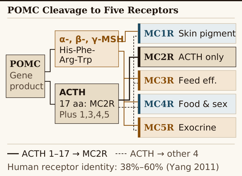
α-, β-, and γ-MSH signal across MC1R, MC3R, MC4R, and MC5R; ACTH stimulates all five receptors and is the sole endogenous agonist for adrenal MC2R, requiring its first 17 amino acids. Sources: Krude (1998), Yang (2011), Cai & Hruby (2016), Yuan & Tao (2022).Why receptor selectivity is chemically difficult
After the first melanocortin receptor genes were cloned by Mountjoy, Cone, and colleagues in 1992, sequence comparisons exposed why synthetic peptides cross between subtypes. Melanocortin receptors are the smallest G protein-coupled receptors known, and human subtypes share 38% to 60% sequence identity—highest between MC4R and MC5R (60%), 45% between MC3R and MC1R or MC2R, and lowest between MC2R and MC4R (38%).
Every natural melanocortin agonist derived from POMC carries the evolutionarily conserved four-amino-acid sequence His-Phe-Arg-Trp. Functional mapping reviewed by Yuan and Tao (2022) distinguishes each receptor's activation requirement: His-Phe-Arg-Trp is the minimal MSH sequence required for MC1R, MC3R, and MC5R activation, three amino acids (Phe-Arg-Trp) suffice in MSH for MC4R, and the first 17 amino acids of ACTH are required for MC2R. Because four pockets recognize that short motif, chemists have faced considerable difficulty developing highly selective ligands.
Receptor-by-receptor accounting: MC1R through MC5R
- MC1R (skin, hair, and immune cells): Located on cutaneous melanocytes—and on leukocytes, where it may mediate anti-inflammatory effects—MC1R governs the balance between red pheomelanin, which can generate free radicals under ultraviolet light, and photoprotective black eumelanin. In 1995, Valverde and colleagues found human
MC1Rsequence variants in over 80% of individuals with red hair, fair skin that tans poorly, or both, compared with fewer than 20% of brown- or black-haired individuals and less than 4% of good tanners. A 2025 review (disclosing editorial assistance funded by Rhythm Pharmaceuticals) counts roughly 200 identified allelic variants with about 60% prevalence among individuals of European ancestry, and notes that MC1R signaling promotes UV-induced DNA nucleotide excision repair independently of melanin pigmentation. - MC2R (adrenal cortex): The lone structural outlier. ACTH is the only known endogenous ligand for MC2R, making it the sole agonist that stimulates all five MCRs, and the first 17 amino acids of ACTH are required for MC2R activation;
α-,β-, andγ-MSHcannot bind or activate MC2R. As Yang (2011) details, substituting phenylalanine-7 with D-phenylalanine (D-Phe7) in ACTH decreases ligand binding affinity and potency at MC2R, whereas substitutingPhe7withD-Phein MSH increases affinity and potency at MC1R, MC3R, MC4R, and MC5R. - MC3R and MC4R (hypothalamus and central nervous system): Mouse knockout studies established nonredundant neural roles: MC3R regulates feed efficiency, while MC4R controls both food intake and energy expenditure. Mice with targeted MC4R inactivation develop maturity-onset obesity with hyperphagia, hyperinsulinemia, and hyperglycemia. A year later, Krude et al. (1998) documented the first two human probands with POMC mutations, defining an endocrine triad of early-onset obesity, adrenal insufficiency, and red hair pigmentation. Central MC4R also participates in brain pathways involved in sexual response.
- MC5R (exocrine glands): Expressed across multiple human peripheral tissues and implicated primarily in exocrine and sebaceous gland secretion in animal targeted-deletion studies.
EC50 potency at human receptors across the switchboard
Pharmacologists compare receptor activity using EC50—the concentration producing half-maximal activation in in vitro assays at human melanocortin receptors, reported in nanomolar (nM), where a lower number means higher potency. In vitro receptor EC50 values do not measure blood-brain-barrier entry, implant release kinetics, or clinical exposure, but they expose the selectivity profile of each peptide.
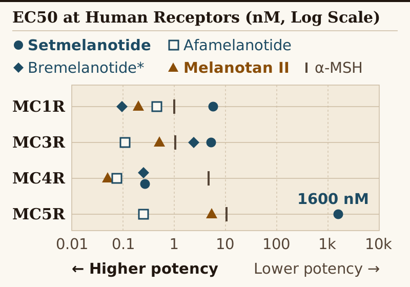
EC50 values toward the left reflect stronger activation at human melanocortin receptors. Data compiled from Table 3 of Yuan & Tao (2022) (primary assay set from Kievit et al., 2013; *bremelanotide from Durek et al., 2018, where MC5R was not reported).| Peptide | MC1R (nM) | MC3R (nM) | MC4R (nM) | MC5R (nM) |
|---|---|---|---|---|
Native α-MSH |
1.01 | 1.04 | 4.7 | 10.5 |
| Afamelanotide (NDP-MSH) | 0.462 | 0.109 | 0.075 | 0.253 |
| Bremelanotide* | 0.095 | 2.4 | 0.25 | Not reported |
| Setmelanotide | 5.8 | 5.3 | 0.27 | 1600 |
| Melanotan II | 0.2 | 0.51 | 0.05 | 5.33 |
Two contrasts stand out in Yuan and Tao's Table 3 (compiling Kievit et al., 2013, and *Durek et al., 2018 for bremelanotide). First, Melanotan II acts as a nonselective agonist with nanomolar to subnanomolar potency across MC1R (0.2 nM), MC3R (0.51 nM), MC4R (0.05 nM), and MC5R (5.33 nM). Second, even linear afamelanotide shows subnanomolar potency at MC3R and MC4R in human-receptor assays—underscoring why clinical development relies on formulation control and tissue access as well as receptor affinity.
Natural brakes, allosteric pockets, and biased signaling
Unlike many hormone receptors, melanocortins exhibit high basal activity. As King and colleagues note, MCRs 1, 3, 4, and 5 maintain high constitutive (ligand-independent) activity, allowing antagonists to decrease basal cyclic AMP levels. Two endogenous proteins fill that role: agouti signaling protein (ASIP) selectively antagonizes MC1R and MC4R, while agouti-related protein (AgRP) selectively antagonizes MC3R and MC4R.
As Yingkui Yang (2011) defines the binding geometry, endogenous agonists bind the orthosteric site—the receptor area where natural ligands dock—whereas an allosteric site is distinct from that endogenous binding site. Modern MC4R drug design also examines biased signaling: MC4R can couple to three major G-protein families (Gs, Gi/o, and Gq). Earlier clinical MC4R obesity candidates failed in human trials despite preclinical efficacy due to poor efficacy and side effects. By contrast, setmelanotide potently activates MC4R Gαq signaling that AgRP cannot efficiently antagonize—a pathway linked in preclinical studies to reduced food intake without cardiovascular pressor effects.